Where Is Ivc Filter Placed
Placement of an junior vena cava (IVC) filter may be a required for a deep vein thrombosis (DVT). Vena cava filters may exist temporary or permanent; the decision is based on an individualized basis. These filters are reserved for patients who are unable to accept blood-thinning medications or for those at loftier risk for developing recurrent DVT with pulmonary embolism (PE).
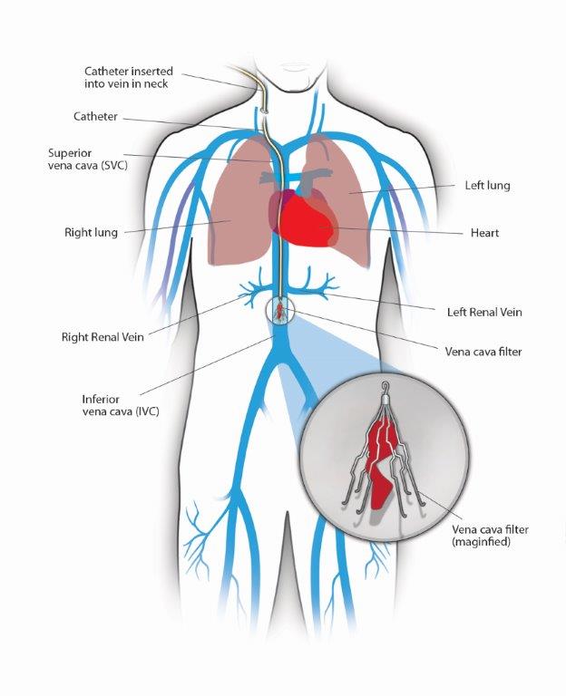
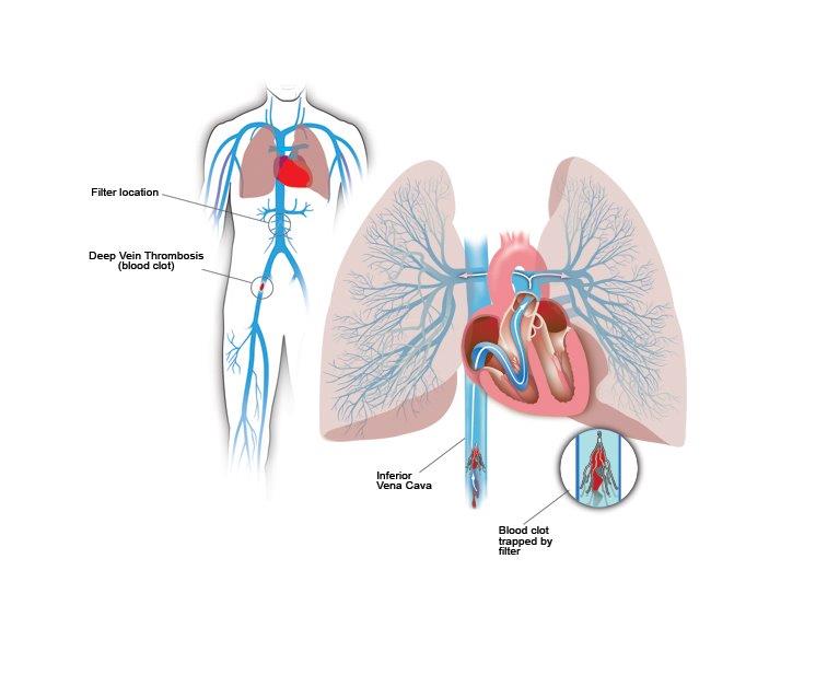
The junior vena cava is a large vein in your abdomen that transports blood dorsum to your heart from the lower extremities. IVC filters are small umbrella-shaped wire devices placed in the inferior vena cava. The filters are designed to trap claret clots from travelling from your legs to the lungs.

The procedure is done using x-ray in the operating room or in the interventional radiology suite. A catheter (tube) is placed from the groin or the neck in the IVC and contrast is used to image the IVC. Through this catheter the filter is placed in the IVC under x-ray guidance.
Rare complications include haemorrhage from the puncture site, reaction to contrast dye, infection at puncture site, small-scale run a risk of pulmonary emboli.

What to Look
After the process your vital signs and the puncture site will exist monitored for a few hours. You can become home the same day with instructions on your blood thinners.
Minimal bruising and discomfort may exist in the puncture site. Whether your surgeon has placed a temporary or permanent IVC filter, information technology is important that you follow upwards routinely.
Removal of IVC filter:
In some situation it may be recommend that the filter be removed. The filter may exist removed if you are able to have blood-thinning medications or if you are no longer considered at high risk of PE.
Sometimes, your team will decide to get out the filter in permanently if you are unable to have claret thinners or the filter has been in a long fourth dimension.
Removing a vena cava filter is usually a simple procedure and is washed as an outpatient procedure. IVC filters are removed through a vein in the cervix. A small catheter (tube) is placed and with guidance of contrast dye and x-ray images, the filter is collapsed inside the catheter and removed through the same puncture pigsty. Afterwards, the catheter is removed and a pressure dressing volition be applied to the puncture site at the finish of the process. The procedure itself usually takes less than an 60 minutes.
Rare complications include bleeding from puncture site, reaction to contrast dye, infection at puncture site, small risk of pulmonary emboli. Sometimes, the filter is embedded inside the vein wall and cannot be removed.
Where Is Ivc Filter Placed,
Source: https://vein.stonybrookmedicine.edu/treatments/inferior-vena-cava-filters
Posted by: vallierekeisheiled.blogspot.com


0 Response to "Where Is Ivc Filter Placed"
Post a Comment